Furfuragaricus microsporusR.L. Zhao & J.X. Li, sp. nov.Fig. 15
Chinese name: 小孢鳞柄蘑菇 (Pinyin: xiǎo bāo lín bǐngmó gū)
Fungal Names registration: FN 572560
Etymology: derived from “micro-”- (small) and “spora”(spore), referring to the small size of the basidiospores pro-duced by this fungus.
Type: China, Hunan Province, Zhangjiajie City, JishouUniversity Zhangjiajie Campus, 29 Jun. 2019, W.Q. Qin,ZRL20190126 (holotype HMAS 282444).
Diagnosis: Furfuragaricus microsporus is characterizedby small-sized basidioma with appressed, furfuraceous,brown to reddish-brown squamules on the pileus. The stipesurface is densely covered with fibrillose-squamulose tosmall scale-like fibrils, concolorous with the pileus. Thebasidiospores are tiny, 3.3–3.9 × 2.3–2.8 μm, ellipsoid tobroadly ellipsoid with small apiculus, hyaline in 5% KOH,thick-walled. Cheilocystidia abundant, clavate to broadlyclavate, with fine crystals at apex.
Macroscopic description: Basidioma small-sized.Pileus about 30 mm, plano-convex to convex when mature,developing furfuraceous depressed squamules of variousbrown on the pileus, pale red, brown, reddish brown, yel-lowish brown, dark brown; densely and darker at center,becoming lighter towards margin, elsewhere (fibrillose)-squamulose; margin appendiculate, often with laceratedvelar remnants that are concolorous with the squamules;context thin, white to off white, yellowish white; no colorchange after damaged; Lamellae free, close to crowded,about 2 mm in width, depressed around the stipe, mostlyventricose in the middle, pale yellowish, yellowish whiteto yellowish grey, grayish to grayish brown, pale brownish,with 2–3 tiers of lamellulae, not changing color after dam-age, becoming dark brown to blackish when dried, marginentire to slightly erose. Stipe c. 55 mm long, 3–5 mm wide,some tapering downwards to base, cylindrical to subcylin-drical, hollow, not discoloring when bruised, developing awhitish to pale reddish background; surface densely fibril-lose-squamulose or covered with small scale-like fibrilswhich are concolorous with those on pileus. Annulus notwell-defined. Odor and taste unknown. Spore prints white.
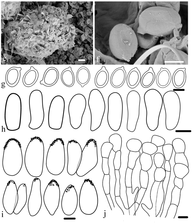
Microscopic description: Basidiospores[55/1/1], (3.1)3.3–3.9(4.6) × (2.0)2.3–2.8(3.0) μm,X = 3.6 ± 0.3 × 2.5 ± 0.3 µm, Q = 1.3–1.5, Qm = 1.4 ± 0.1,most ellipsoid to broadly ellipsoid, thick-walled, smooth,almost hyaline in both water and 5% KOH, with a smallapiculus, without germ pore. Basidia not observed. Basidi-oles abundant, 6.7–10.2 × 3.3–4.7 μm, clavate to broadlyclavate, thin-walled, hyaline with thin walls. Cheilocyst-idia 12.5–16.7 × 6.1–8.2 µm, abundant, clavate to broadlyclavate, with fine crystals at apex, hyaline with thin walls.Pileus covering an irregular epithelium; consisting ofoblong to short clavate elements, or irregular globose tosubglobose elements, slightly thick-walled, encrusted,often containing pale brownish internal pigment. Clampconnections absent.
Habitat and distribution: terrestrial; solitarily on soil indeciduous broadleaf forests; collected beneath Castaneaat the type locality; currently, known only from HunanProvince, China.
Notes: Phylogenetic analyses based on four loci (ITS,nrLSU, rpb2, and tef1) indicate that Fu. microsporusforms a distinct lineage (Fig. 4). Four other genera—Eriocybe, Mystagaricus, Pseudolepiota, and Xanthagari-cus—are closely related to Furfuragaricus. Eriocybe isdistinguished by a white, woolly, felt-like covering on thepileus and stipe (Vellinga et al. 2011). Mystagaricus isdistinguished by a purplish brown to vinaceous, stronglywoolly, felty pileus surface (Radnóti et al. 2025). Pseu-dolepiota is characterized by a pileus diameter that isequal to or slightly larger than the stipe length. Its pileusis covered with violet-brown to dark ruby squamules, withdull red fibrils between them, and has a white appendicu-late margin (Ge and Yang 2017; Sysouphanthong et al.2021). Xanthagaricus differs in having slenderer basidi-oma than Furfurastipe (Yang et al. 2024a). In addition, thebasidiospores in Furfuragaricus are thick-walled (Fig. 15).The distinct phylogenetic placement of Fu. microsporus,together with its unique combination of macro- and micro-morphological characteristics, supports its recognition asa novel taxonomic entity.
参考翻译:
小孢鳞柄蘑菇
鉴别特征: 小孢鳞柄蘑菇的主要鉴别特征为:子实体小型;菌盖表面具贴伏、糠麸状、褐色至红褐色的鳞片;菌柄表面密被纤维状-鳞片状至小鳞片状纤维,颜色与菌盖相同。担孢子微小,3.3–3.9 × 2.3–2.8 μm,椭圆形至阔椭圆形,具小的尖突,在5% KOH溶液中透明,壁厚。褶缘囊状体丰富,棍棒状至阔棍棒状,顶端具细小的结晶体。
宏观描述: 子实体小型。菌盖直径约30 mm,成熟时平凸至凸形,表面形成糠麸状、下陷的褐色系列鳞片,包括淡红色、褐色、红褐色、黄褐色、暗褐色等;菌盖中央区域密集且颜色较深,向边缘逐渐变淡,其余部分(纤维状)-鳞片状;菌盖边缘具残幕附着,常有撕裂状的菌幕残余,颜色与鳞片同色;菌肉薄,白色至灰白色、黄白色;受伤后不变色。菌离生,密至非常密,宽约2 mm,菌柄周围处下陷,中部多呈腹鼓状,颜色为淡黄色、黄白色至黄灰色、灰色至灰褐色、淡褐色;有小菌褶2–3层;受伤后不变色,干燥后呈暗褐色至近黑色;菌边缘全缘至稍呈啮蚀状。菌柄长约55 mm,宽3–5 mm,部分向下渐细至基部,圆柱形至近圆柱形,中空,受伤后不变色,具白色至淡红色的底色;表面密被纤维状-鳞片状或覆盖小鳞片状纤维,颜色与菌盖上的相同。菌环不明显。气味与味道未知。孢子印白色。
显微描述: 担孢子 [55/1/1],(3.1)3.3–3.9(4.6) × (2.0)2.3–2.8(3.0) μm,平均X = 3.6 ± 0.3 × 2.5 ± 0.3 µm,长宽比Q = 1.3–1.5,平均Qm = 1.4 ± 0.1,大多数为椭圆形至阔椭圆形,壁厚,光滑,在清水和5% KOH中几乎透明,具小尖突,无芽孔。担子未观察到。幼担子丰富,6.7–10.2 × 3.3–4.7 μm,棍棒状至阔棍棒状,薄壁,透明。褶缘囊状体 12.5–16.7 × 6.1–8.2 µm,丰富,棍棒状至阔棍棒状,顶端具细小结晶体,薄壁,透明。菌盖皮层为不规则的上皮状;由长圆形至短棍棒状细胞,或不规则球形至近球形细胞组成,壁稍厚,具结晶体,常含有淡褐色的内部色素。无锁状联合。
生境与分布: 地生;单个或散生于落叶阔叶林中的土壤上;模式产地采自栗属树下;目前仅知分布于中国湖南省。
讨论: 基于四个基因片段(ITS、nrLSU、rpb2、tef1)的系统发育分析表明,小孢鳞柄蘑菇形成了一个独立的谱系(图4)。另外四个属——
Eriocybe、
Mystagaricus、假环柄菇属和黄蘑菇属——与鳞柄蘑菇属关系密切。
Eriocybe的鉴别特征在于菌盖和菌柄具有白色、羊毛状、毡状的覆盖物(Vellinga等 2011)。
Mystagaricus的鉴别特征在于菌盖表面呈紫褐色至酒红色,具强烈的羊毛状、毡毛状覆盖(Radnóti等 2025)。假环柄菇属的特征是菌盖直径等于或略大于菌柄长度,菌盖覆盖有紫褐色至暗红宝石色的鳞片,鳞片间夹杂着暗红色的纤维,且菌盖边缘菌幕残片白色(Ge和Yang 2017;Sysouphanthong等 2021)。黄蘑菇属与鳞柄蘑菇属的区别在于其子实体更为细长(Yang等 2024a)。此外,鳞柄蘑菇属的担孢子为厚壁(图15)。小孢鳞柄蘑菇独特的系统发育位置,加之其宏观与微观形态特征的独特组合,支持将其认定为一个新的分类实体。